
Sunday, September 28, 2025
Contouring for Genitourinary Cancer
Faculty
- Hong Zhang, MD, PhD - Professor of Radiation Oncology, University of Rochester Medical Center,Wilmot Cancer Institute
- Neil Taunk, MD, MS - Assistant Professor of Radiation Oncology and Radiology, University of Pennsylvania, Abramson Cancer Center
Review the case description and images below.
Case Description
- Patient: 65-year-old male
- Initial Diagnosis:
- PSA prior to surgery: 8.6
- Pathologic stage: pT3aN0cM0, Gleason grade group 4
- surgical margins negative, extraprostatic extension at the bladder neck
- Postoperative Course:
- PSA nadir: 0.07 ng/mL
- PSA progression: Increased to 0.7 ng/ml, doubling time 6 months
- Restaging Studies:
- PSMA PET: No evidence of PSMA-avid disease
- Multiparametric MRI: No evidence of disease
- Current Management:
- Initiated on salvage radiation therapy with 6 months of ADT
Question
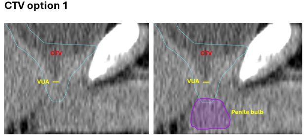
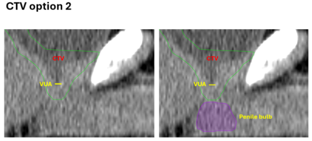
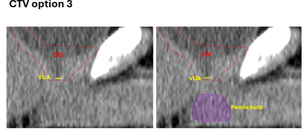
Which of the displayed sagittal below contours appropriately represents the inferior border of the prostate bed CTV?
Faculty Follow Up
ANSWER - CTV Option 2
Defining the Inferior Border of Prostate Bed CTV
- High incidence of recurrence at the vesicourethral anastomosis (VUA)
- Accurate delineation of the prostate bed clinical target volume (CTV), including the inferior border, is critical to ensure adequate coverage of this high-risk region while respecting normal tissue tolerances
- Consensus guidelines vary in how inferior borders are defined
CTV Inferior Border Contour Guidelines
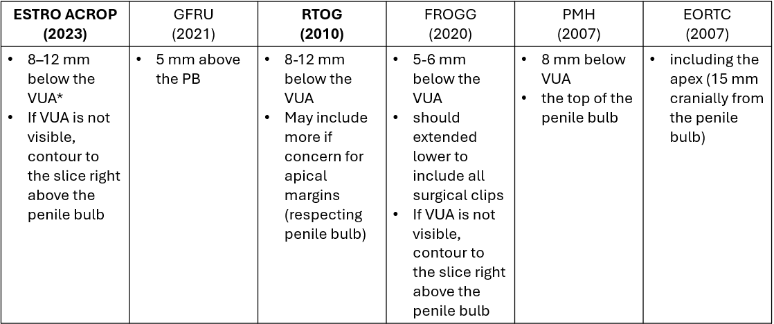
*VUA: vesicourethral anastomosis
MR can reduce inter-observer variability in prostate bed radiotherapy planning
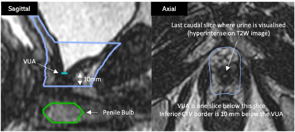
- MR sim or planning MR
- Identify the last slice on the axial view where urine is visualized, VUA is one slice below this slice
- Using the sagittal view to confirm the location of VUA
- High incidence of recurrence at the vesicourethral anastomosis (VUA)
- Accurate delineation of the prostate bed clinical target volume (CTV) is critical to ensure adequate coverage of this high-risk region while respecting normal tissue tolerances
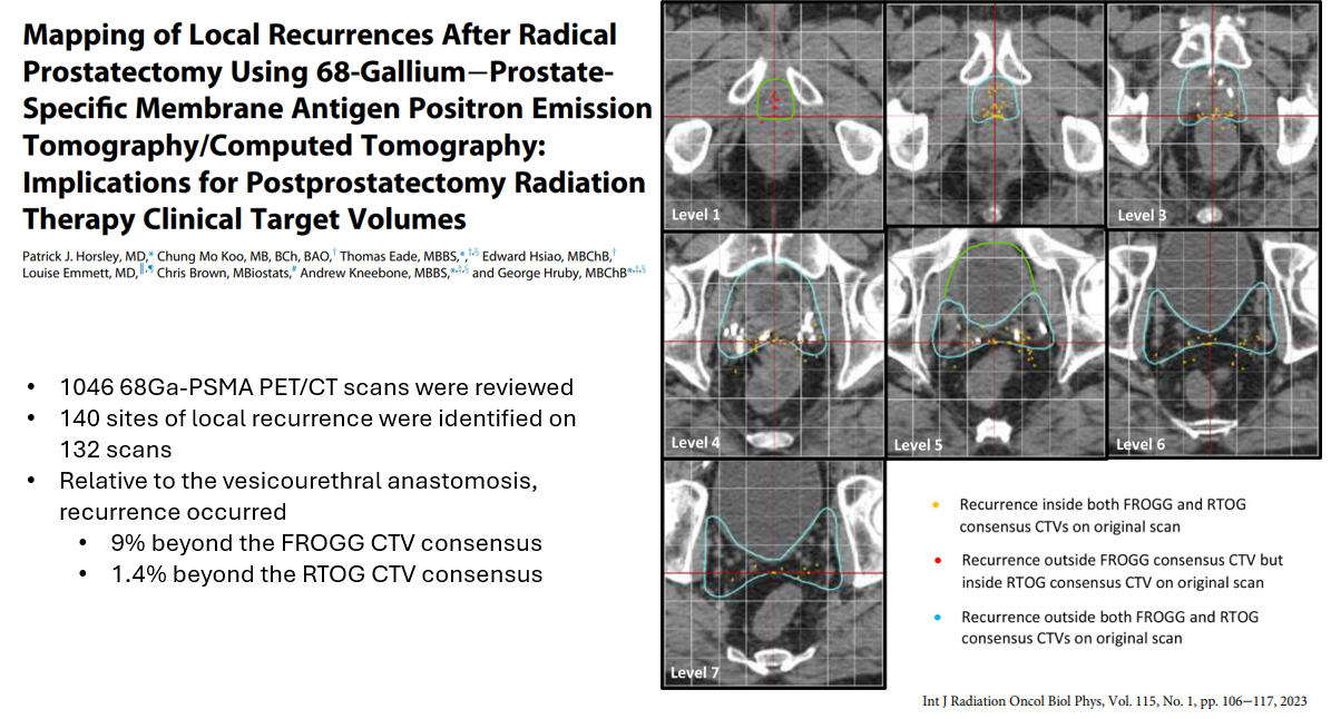
- Integration of modern imaging, MRI prostate and PSMA PET, for treatment planning
The best way to define the inferior border of CTV
Patient-tailored approach
Using imaging and clinical context to guide individualized adjustments to the inferior border of CTV.