
Monday, September 29, 2025
Contouring for Central Nervous System Cancer
Faculty:
- Michelle Kim, MD - Associate Chair, Department of Radiation Oncology, Clinical Professor of Radiation Oncology Medical School, University of Michigan
- Helen Shih, MD, MS, FASTRO, MPH, AM - Medical Director, MGH Proton Therapy Centers, Massachusetts General Hospital
Review the case description and images below.
Case Description
A 45 year old right-handed male presented with headaches, nausea, double vision and word-finding difficulties. MRI reveals hydrocephalus and 1 cm of midline shift from a large, heterogeneous left frontal mass that is predominantly non-enhancing, with patchy areas of enhancement. The patient undergoes awake craniotomy and subtotal resection with pathology revealing Astrocytoma, IDH-mutant, CNS WHO grade 3 with MGMT promoter methylation and without CDKN2A/B homozygous deletion.
Which clinical target volume contour is the most appropriate?
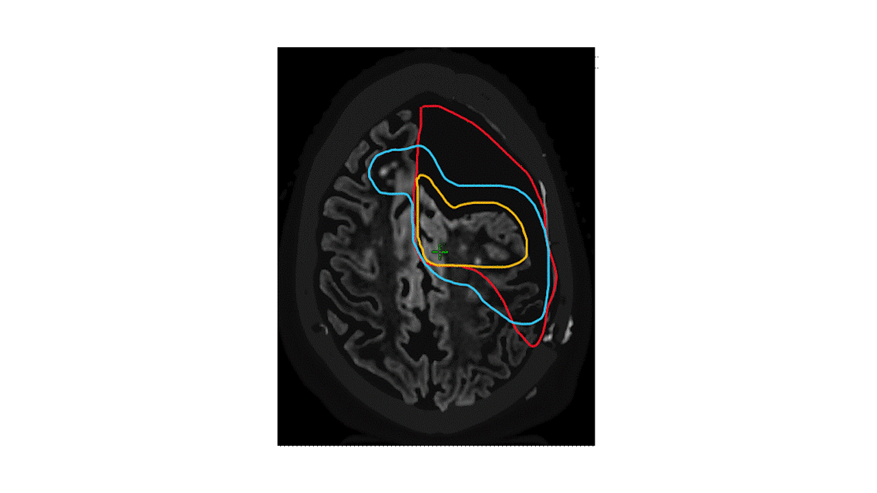
A. Surgical cavity + residual T1-post contrast enhancement and an anatomically constrained 1.5 cm CTV margin
B. Tumor bed + residual T1-post contrast enhancement + 2 cm CTV margin including ventriculostomy tract
C. Tumor bed + residual T1-post contrast enhancement + residual T2/FLAIR and an anatomically constrained 1.5 cm CTV margin
Faculty Follow Up
ANSWER - C
Imaging Tips
- Immediate preoperative and a recent postoperative MRI are needed to best define glioma target volumes. Preoperative imaging is invaluable in defining the full extent of tumor and its margins that may not be apparent after the tumor resection and helps to locate the true tumor bed after any anatomical shifts.
- Gliomas are infiltrative tumors that extend through the brain parenchyma beyond what may be visibly apparent on T1-post contrast and T2/FLAIR MR images. Both imaging sequences are contributory to defining the radiographically detectable disease.
How We Do It
Defining GTV:
- Both T1 post contrast and T2/FLAIR signals represent parts of the tumor. Postoperative T2/FLAIR is most representative of the residual lower grade glioma that only variably enhances and is best defined using high resolution (3D) FLAIR images.
- However, not every T2-hyperintense lesion is part of the tumor, and chronic T2-hyperintensities representing cerebrovascular sequelae or other non-tumor related changes such as from a ventriculostomy tract separate from tumor should not be considered part of the GTV (e.g. incorrect option B (blue)).
- The edge of tumor bed is included in the residual GTV that is subsequently expanded.
Defining CTV: Don’t overextend
- Anatomical barriers exist such as the falx/dura and sulci which will ordinarily prevent direct tumor extension into neighboring brain tissue.
- Uniform expansion of 1-1.5 cm is most commonly applied with subsequent editing to remove CTV extension at anatomical barriers. The CTV may be restricted to the supratentorium in most cases, and out of adjacent uninvolved OAR’s such as the chiasm.
- Large surgical cavities that predominantly consist of CSF (e.g. following lobectomy without circumferential brain parenchyma) do not need to be treated. It is highly unlikely that tumor will regrow in that space without brain parenchymal infrastructure. This helps to avoid alopecia and contribution to fatigue from treatment (e.g. incorrect option A (red)).
Defining CTV: Don’t underextend
- On the other hand, sufficient CTV margin along possible areas of white matter spread is important. Both options A (red) and B (blue) exemplify insufficient CTV margin beyond the non-enhancing disease already present in the corpus callosum.
- It is important to be mindful that the 3-dimensional extension of tumor cells can spread around barriers off plane from a given image. The entire scan should be viewed in all planes to appreciate the 3-dimensional growth pattern.
CTV Pro tips:
- Small extension of the CTV into CSF space of surgical cavities or ventricles can help to prevent underdosing if there are further anatomical shifts during treatment, such as from reactive swelling or regression of the cavity. Large shifts should prompt adaptive replanning.
- Gliomas travel more easily along deep white matter bundles such that larger CTV margins may be favored here as compared to the more peripheral gray matter.